Dr. Rahul Chauhan
Specialist in shoulder, knee, spine endoscopy and ultrasound guided orthopedic procedures.
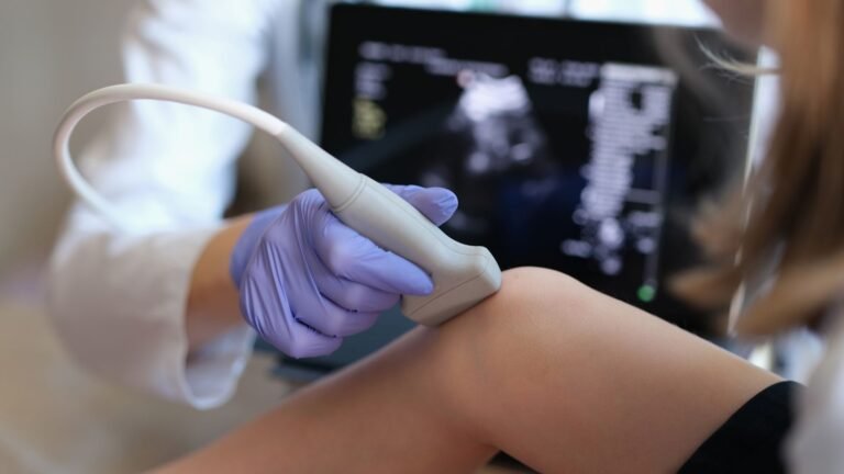
The Posterior Cruciate Ligament (PCL) is one of the key stabilizing ligaments of the knee. When it tears due to trauma, patients may experience deep knee pain, difficulty walking downhill or downstairs, and a feeling of the knee “giving way.”
If the ligament does not heal properly, arthroscopic PCL repair or reconstruction can restore stability and protect the knee from long-term damage.
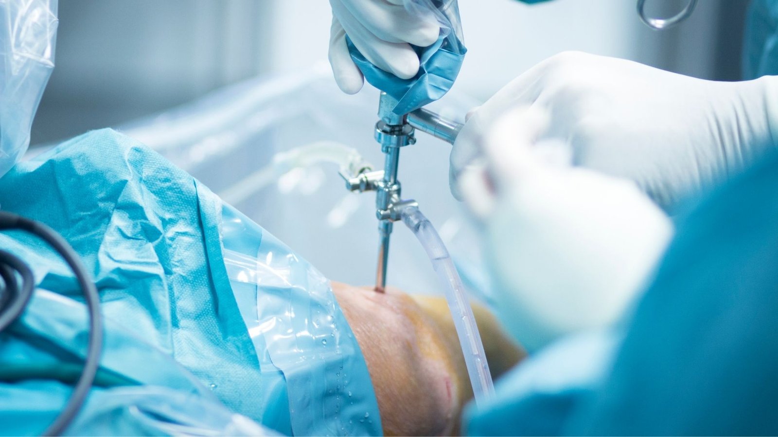
PCL surgery is performed using keyhole (arthroscopic) techniques, which means only tiny incisions are made around the knee. A high-definition camera and fine instruments are inserted to repair or reconstruct the torn ligament with precision.
Depending on the type and location of the tear
If the ligament has peeled off cleanly from its attachment and tissue quality is good, the surgeon may reattach it using sutures and anchors.
More commonly, the torn PCL needs to be replaced with a graft, which acts as a new ligament. Grafts are usually taken from:
The graft is placed exactly where the original PCL was, restoring normal knee biomechanics.
Surgery is recommended for patients who have:
Early surgical stabilization helps prevent meniscus damage and early arthritis.
Compared to older open procedures, the arthroscopic technique offers:
The goal is not just to stop instability but to restore confidence, function and knee longevity.
A simplified overview:
Patients usually begin walking with support the same or next day, depending on protocol.
Healing takes time because the PCL deep inside the knee requires gradual graft integration.
| Time After Surgery | Progress |
|---|---|
| First 6 weeks | Brace support, assisted walking, early therapy |
| 6–12 weeks | Improved mobility & strengthening |
| 3–6 months | Functional training and light jogging |
| 6–9 months | Sports-specific training |
| 9–12 months | Return to pivoting / contact sports (only after clearance) |
A structured physiotherapy program is essential for the best results.
Most patients return to:
High-level athletes can also return to competitive sport with proper rehabilitation.
Arthroscopic PCL repair or reconstruction is a highly effective and minimally invasive solution for PCL injuries that do not heal on their own. By restoring knee stability early, the procedure protects the joint from future wear-and-tear and helps patients regain an active, confident and pain-free life.